Home
/ Back Muscle Chart - The Muscular System Anatomical Chart Muscle Anatomy Human Body Png Clipart Abdomen Anatomy Back Cardiac Muscle - The back's muscles start at the top of the back (named the cervical vertebrae) and go to the tailbone (also named the coccyx).
Back Muscle Chart - The Muscular System Anatomical Chart Muscle Anatomy Human Body Png Clipart Abdomen Anatomy Back Cardiac Muscle - The back's muscles start at the top of the back (named the cervical vertebrae) and go to the tailbone (also named the coccyx).
Back Muscle Chart - The Muscular System Anatomical Chart Muscle Anatomy Human Body Png Clipart Abdomen Anatomy Back Cardiac Muscle - The back's muscles start at the top of the back (named the cervical vertebrae) and go to the tailbone (also named the coccyx).. We've created a free trigger point chart, which includes fybromyalgia treatment and reflexology information. Muscle strain is often the cause of back pain from heavy lifting or vigorous exercise. The trapezius and latissimus dorsi muscles connect the upper limb to the vertebral column. For images of the muscle, click on each link under location. There are three different muscle groups found in the back:
Superficial back muscles, intermediate back muscles and intrinsic back muscles.the intrinsic muscles are named as such because their embryological development begins in the back, oppose to the superficial and intermediate back muscles which develop elsewhere and are therefore classed as extrinsic muscles. Leaning back to straight vertical and all points in between. Deep back muscles diagram the superficial layer contains the splenius cervicis and splenius capitis muscles. Certain back muscles extend to other areas, like the shoulders, upper arms, and thighs. The muscles of the lower back help stabilize, rotate, flex, and extend the spinal column, which is a bony tower of 24 vertebrae that gives the body structure and houses the spinal cord.
Skeletal Muscle Wikipedia from upload.wikimedia.org Loss of control of the bowel or bladder and retention of urine may. The most common type of back pain is muscle pain—also called muscle strain or soft tissue strain. Other muscles are small and cover much less space. For more anatomy content please follow us and visit our website: Lower back muscle diagram anatomy does degenerative disc disease affect the lower back muscle? Extends spine and trunk back. Certain back muscles extend to other areas, like the shoulders, upper arms, and thighs. When back development is the goal, stick to one of these variations.
The back isn't only one of the body's biggest and strongest body parts, it's also the most complicated in terms of being a series of interconnected muscle groups.
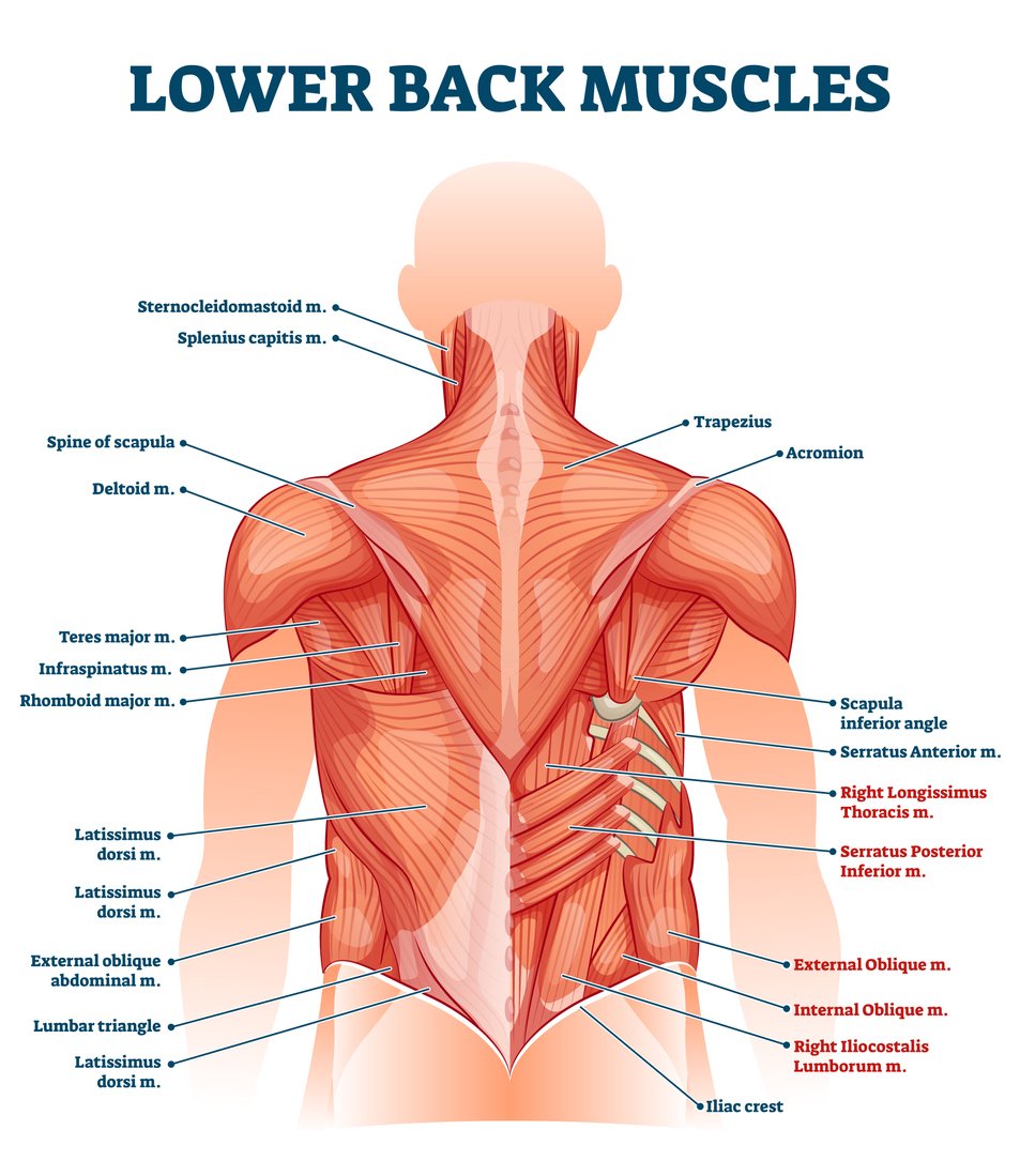
The vast majority of back problems improve on their own or with nonsurgical treatment. What is the origin and insertion of the rhomboid minor and major muscle? The deltoid, teres major, teres minor, infraspinatus, supraspinatus (not shown) and subscapularis muscles (not shown) all extend from the scapula to the humerus and act on the shoulder joint. Both the deltoid and the trapezius are firmly attached to the spine of the scapula. Some of these muscles are quite large and cover broad areas. Muscles found in the superficial group include rhomboid major, rhomboid minor, levator scapulae, trapezius, latissimus dorsi. There are three different muscle groups found in the back: We hope this picture anatomy of back muscles diagram can help you study and research. The intermediate layer contains the erector spinae muscles, whose many functions include the extension and lateral flexion of the spine, head and neck. The deep back muscles, also called intrinsic or true back muscles, consist of four layers of muscles: The most common type of back pain is muscle pain—also called muscle strain or soft tissue strain. Anatomy chart courtesy of fcit the latissimus dorsi muscles (also known as the lats) are the largest muscles of the back. The rhomboid muscle is activated as you bring and squeeze your scapula or shoulder blades back and together.
Related posts of muscles of the lower back and hip diagram muscle anatomy of lower back. We've created a free trigger point chart, which includes fybromyalgia treatment and reflexology information. To learn more about the anatomy of the spine, watch this video. We think this is the most useful anatomy picture that you need. See back muscle anatomy stock video clips.
Muscle Chart With German Description Of The Most Important Muscles Of The Human Body Front And Stock Vector Vector And Low Budget Royalty Free Image Pic Esy 046579061 Agefotostock from previews.agefotostock.com This chart shows the outermost layer, called the superficial layer, of our major muscles. This increases blood flow to the muscle normalizing it and bringing it back to a healthy state. Muscles found in the superficial group include rhomboid major, rhomboid minor, levator scapulae, trapezius, latissimus dorsi. The muscles of the back can be arranged into 3 categories based on their location: For images of the muscle, click on each link under location. What is the origin and insertion of the rhomboid minor and major muscle? We've created a free trigger point chart, which includes fybromyalgia treatment and reflexology information. Some of the links in the post above are affiliate links..
We've created a free trigger point chart, which includes fybromyalgia treatment and reflexology information.
Certain back muscles extend to other areas, like the shoulders, upper arms, and thighs. Lower back muscle diagram anatomy does degenerative disc disease affect the lower back muscle? Creatine research more than a sports supplement read more…. Muscle charts of the human body for your reference value these charts show the major superficial and deep muscles of the human body. This is a diagram of the larger and more surface muscles of the low back. To download your free copy click the link. Symptoms of muscle pain include: Brings hip away from body. The deep back muscles, also called intrinsic or true back muscles, consist of four layers of muscles: Claim your free copy of the client back care guide today. Another common cause of lower back and hip pain is disc injury. Anatomy chart courtesy of fcit the latissimus dorsi muscles (also known as the lats) are the largest muscles of the back. The intermediate layer contains the erector spinae muscles, whose many functions include the extension and lateral flexion of the spine, head and neck.
To download your free copy click the link. Loss of control of the bowel or bladder and retention of urine may. To learn more about the anatomy of the spine, watch this video. The trapezius and latissimus dorsi muscles connect the upper limb to the vertebral column. Anatomy chart courtesy of fcit the latissimus dorsi muscles (also known as the lats) are the largest muscles of the back.
Lower Back Muscle Anatomy And Low Back Pain from ix-cdn.b2e5.com Claim your free copy of the client back care guide today. The muscles of the lower back help stabilize, rotate, flex, and extend the spinal column, which is a bony tower of 24 vertebrae that gives the body structure and houses the spinal cord. Muscles are usually work in pairs because although they can contract and shorten (flex), they are pulled by an opposite (antagonist) muscle to straighten out (extend) again. Creatine research more than a sports supplement read more…. We hope this picture anatomy of back muscles diagram can help you study and research. We think this is the most useful anatomy picture that you need. Muscle charts of the human body for your reference value these charts show the major superficial and deep muscles of the human body. Most of the time, back muscle pain is diagnosed then treated with little more than a prescription of rest, painkillers and muscle relaxants.
Muscles found in the superficial group include rhomboid major, rhomboid minor, levator scapulae, trapezius, latissimus dorsi.
Loss of control of the bowel or bladder and retention of urine may. The intermediate layer contains the erector spinae muscles, whose many functions include the extension and lateral flexion of the spine, head and neck. Symptoms of muscle pain include: Muscle anatomy get body smart 12 photos of the muscle anatomy get body smart muscle anatomy get body smart, human muscles, muscle anatomy get body smart. Muscles found in the superficial group include rhomboid major, rhomboid minor, levator scapulae, trapezius, latissimus dorsi. For the purposes of this feature, we're dividing the back into its four main regions: Anatomy chart courtesy of fcit the latissimus dorsi muscles (also known as the lats) are the largest muscles of the back. The deltoid, teres major, teres minor, infraspinatus, supraspinatus (not shown) and subscapularis muscles (not shown) all extend from the scapula to the humerus and act on the shoulder joint. This is a diagram of the larger and more surface muscles of the low back. Muscle anatomy of lower back 12 photos of the muscle anatomy of lower back anatomy muscles lower back hip, anatomy of lower back muscle pain, muscle anatomy in lower back, muscle anatomy of lower back, muscle anatomy of lumbar spine, human muscles. These structures work together to support the body, enable a range of movements, and send messages from the. Most of the time, back muscle pain is diagnosed then treated with little more than a prescription of rest, painkillers and muscle relaxants. This procedure is one of the most powerful yet simple ways to treat muscle pain and discomfort.


 and go to the tailbone (also named the coccyx).)